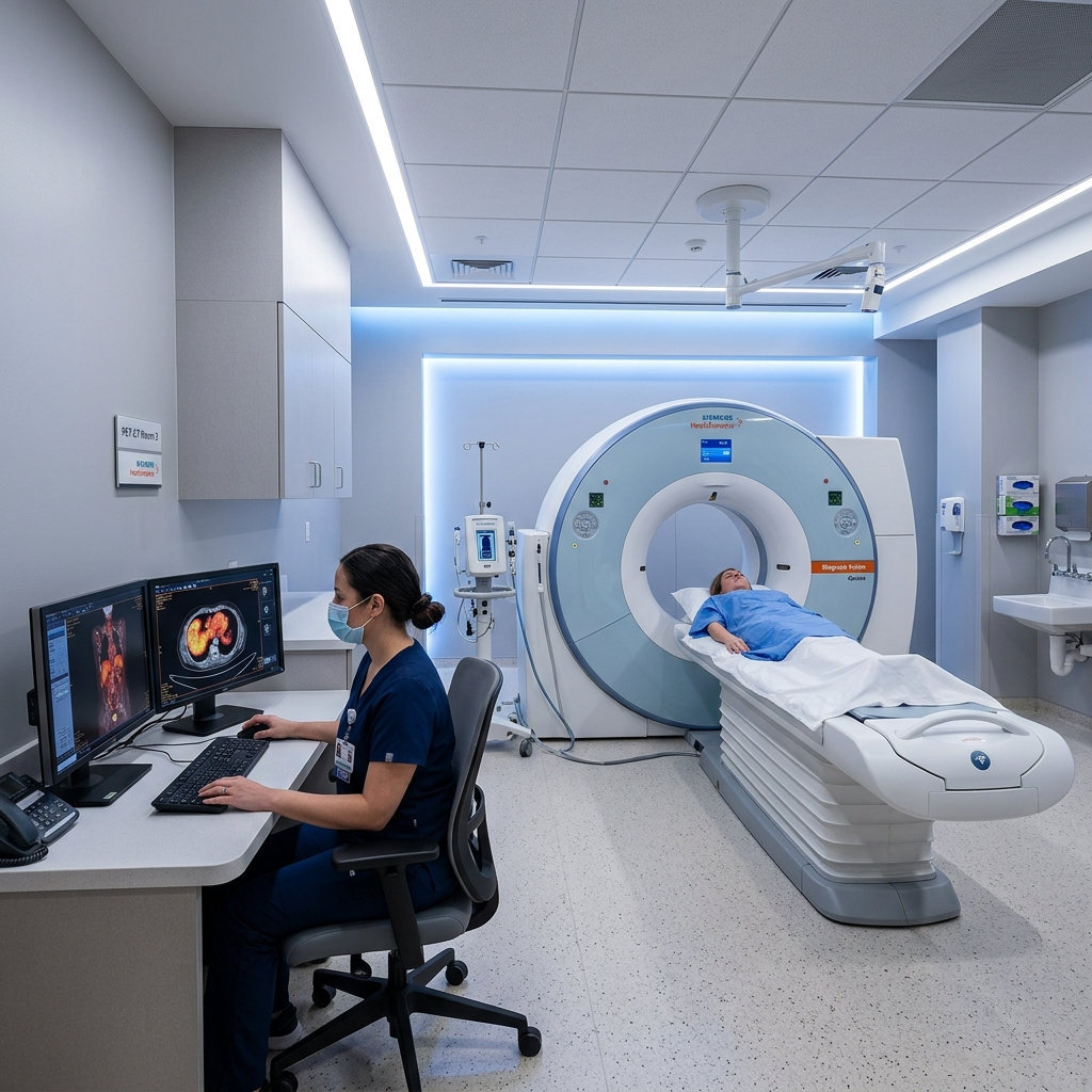
PET-CT imaging suite
Diagnostic Imaging & Technology

Clear answers.
Carefully planned care.

Our imaging team helps patients and specialists understand what is happening, prepare for the next step and make informed care decisions.

Imaging with Purpose

Every examination starts with a clinical question.

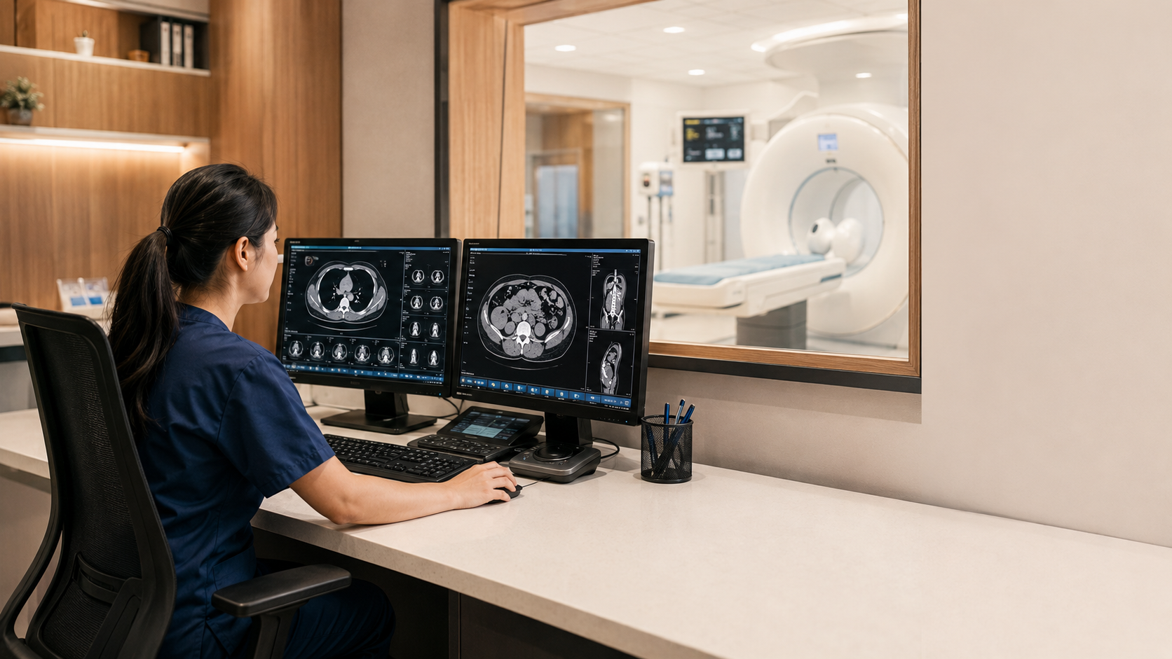
Imaging technologist reviewing diagnostic images in control room

Imaging may support screening, diagnosis, staging, treatment planning or follow-up. The care team selects an examination according to each patient’s clinical needs.

01
Detect & investigate Create detailed views that help clinicians examine a concern.
02
Stage & plan Understand location and extent to support coordinated planning.
03
Guide & monitor Support treatment planning and assessment over time.
How Imaging Supports Care

One connected pathway, centred on the patient.

From the first clinical question to a care decision, each stage provides information the clinical team can use.

01 START

Clinical question

The referral defines what the care team needs to understand.
02 SELECT

Examination

The appropriate modality and protocol are selected.

03 ACQUIRE

Imaging

Images are obtained with attention to safety and quality.

04 INTERPRET

Specialist review

Findings are assessed and communicated through the care pathway.

05 DECIDE

Care decision

The results help inform the appropriate next step.

06 SUPPORT

Cancer Support

Symptom control, comfort, quality of life and continuous patient support.

Scan & Technology Library

Find the guide for your examination.

Each guide explains why an examination may be used, how to prepare, what happens during it and what may follow afterwards.
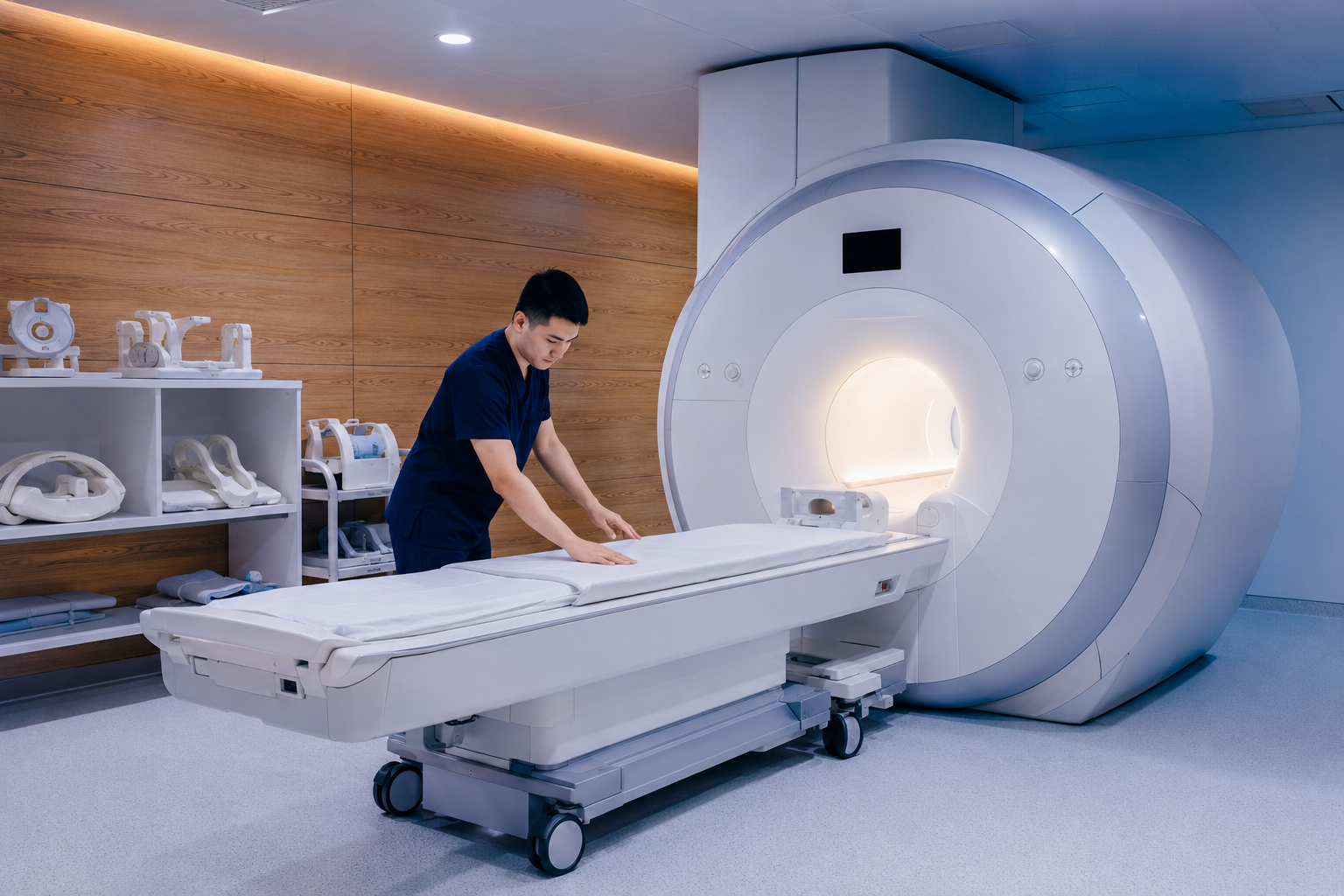
Magnetic Resonance Imaging (MRI) environment
01 Cross-sectional imaging

Magnetic Resonance Imaging (MRI)

Detailed soft-tissue imaging created without ionising radiation.

PET-CT Scan environment
02 Molecular & anatomical imaging

PET-CT Scan

Functional information shown together with detailed anatomical imaging.
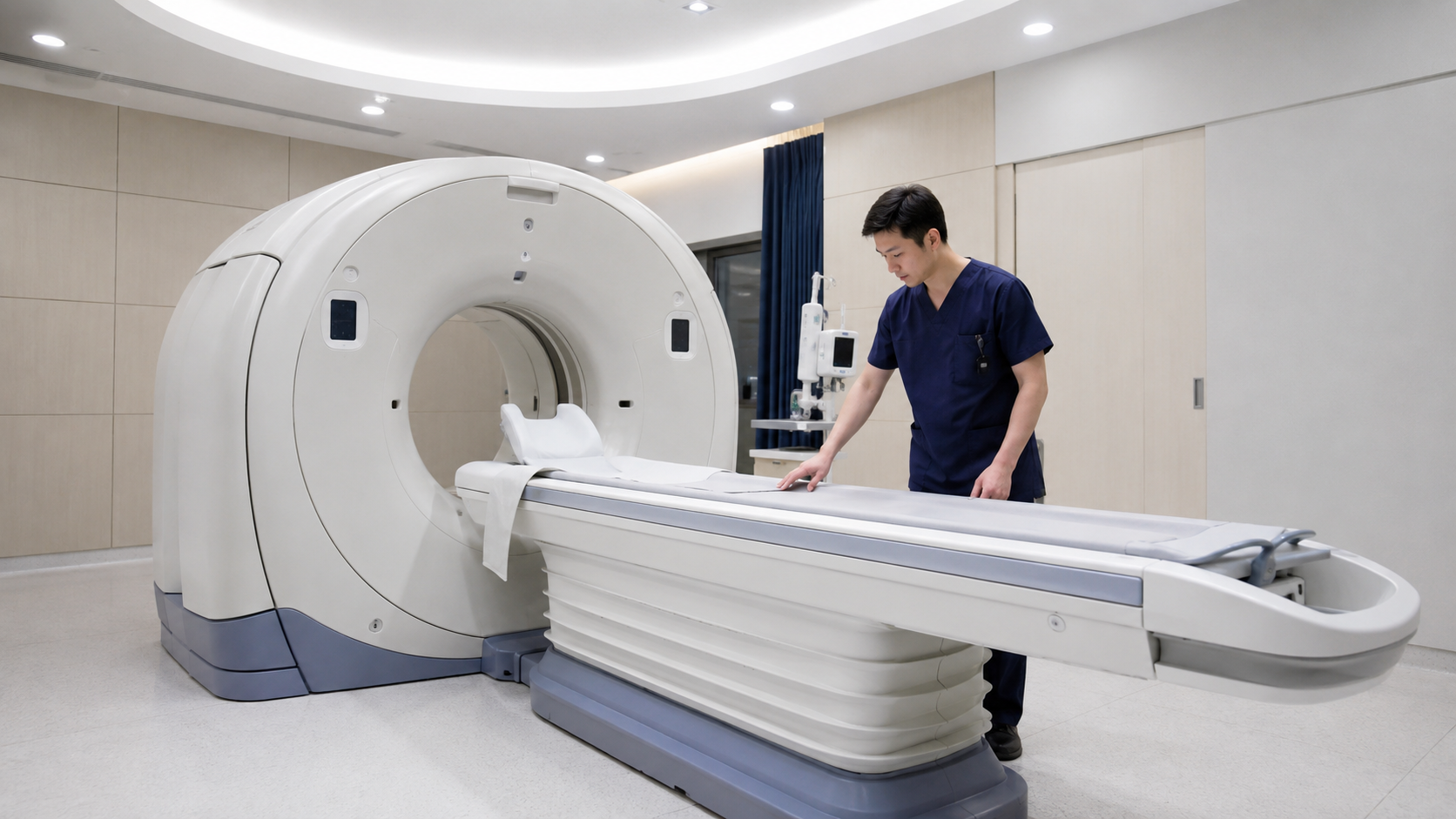
Computed Tomography (CT) environment
03 Fast anatomical imaging

Computed Tomography (CT)

Cross-sectional imaging that may support assessment, planning and follow-up.
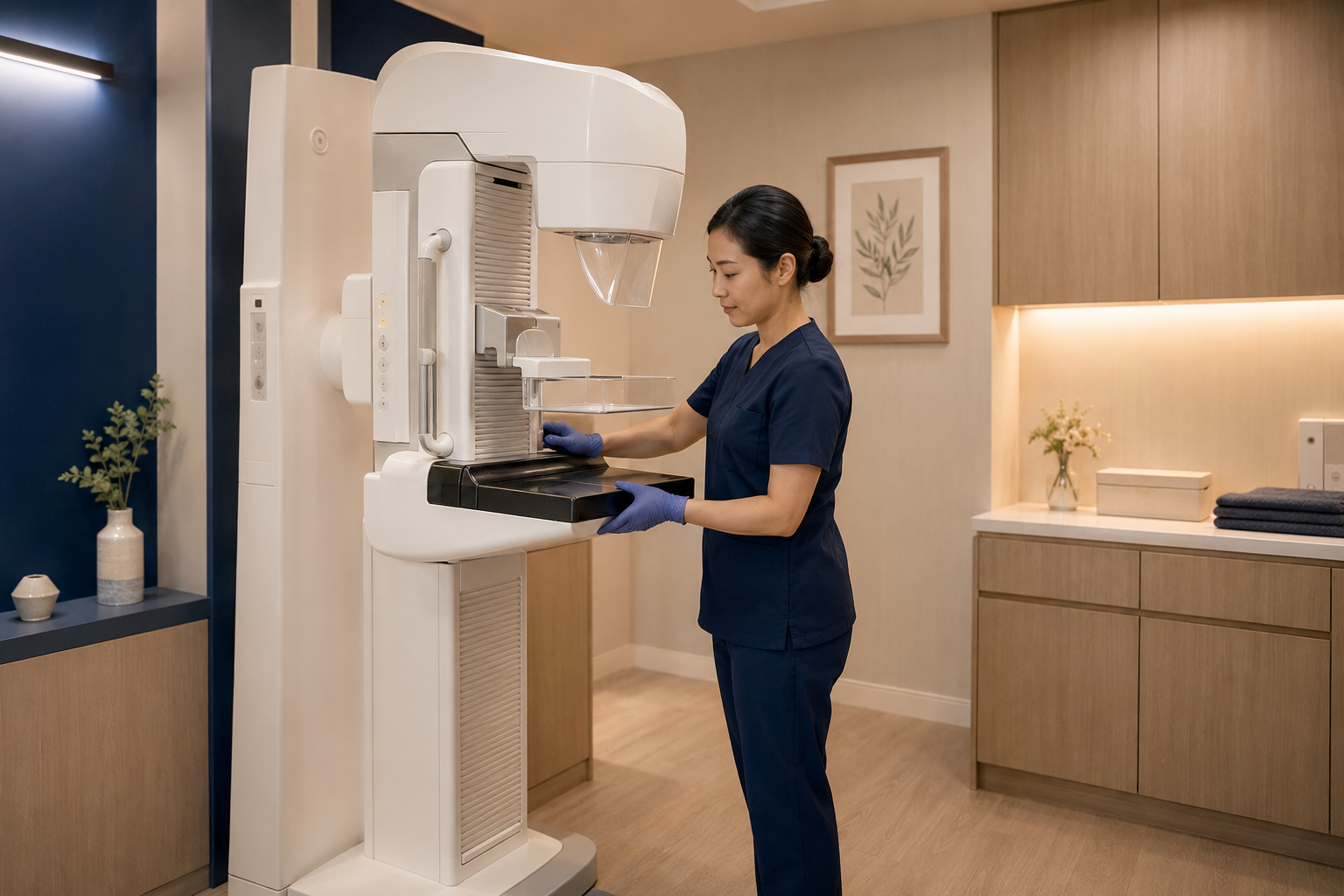
Mammogram environment
04 Breast imaging

Mammogram

Breast imaging for screening or diagnostic assessment where appropriate.
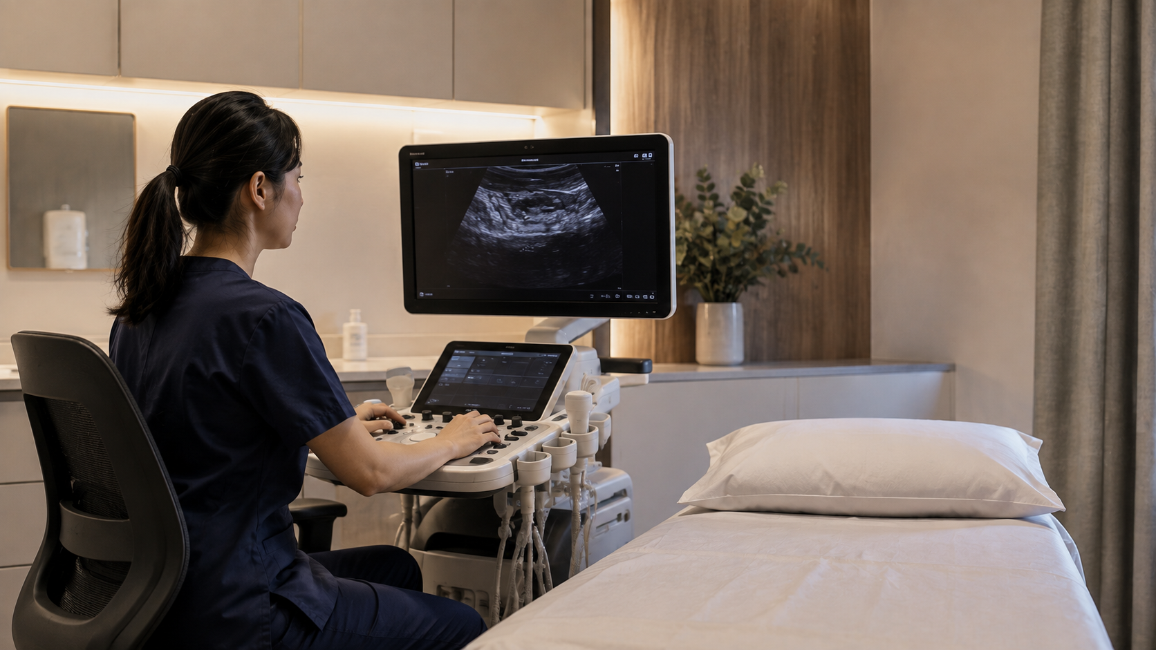
Ultrasound environment
05 Ultrasound

Real-time imaging using sound waves rather than ionising radiation.
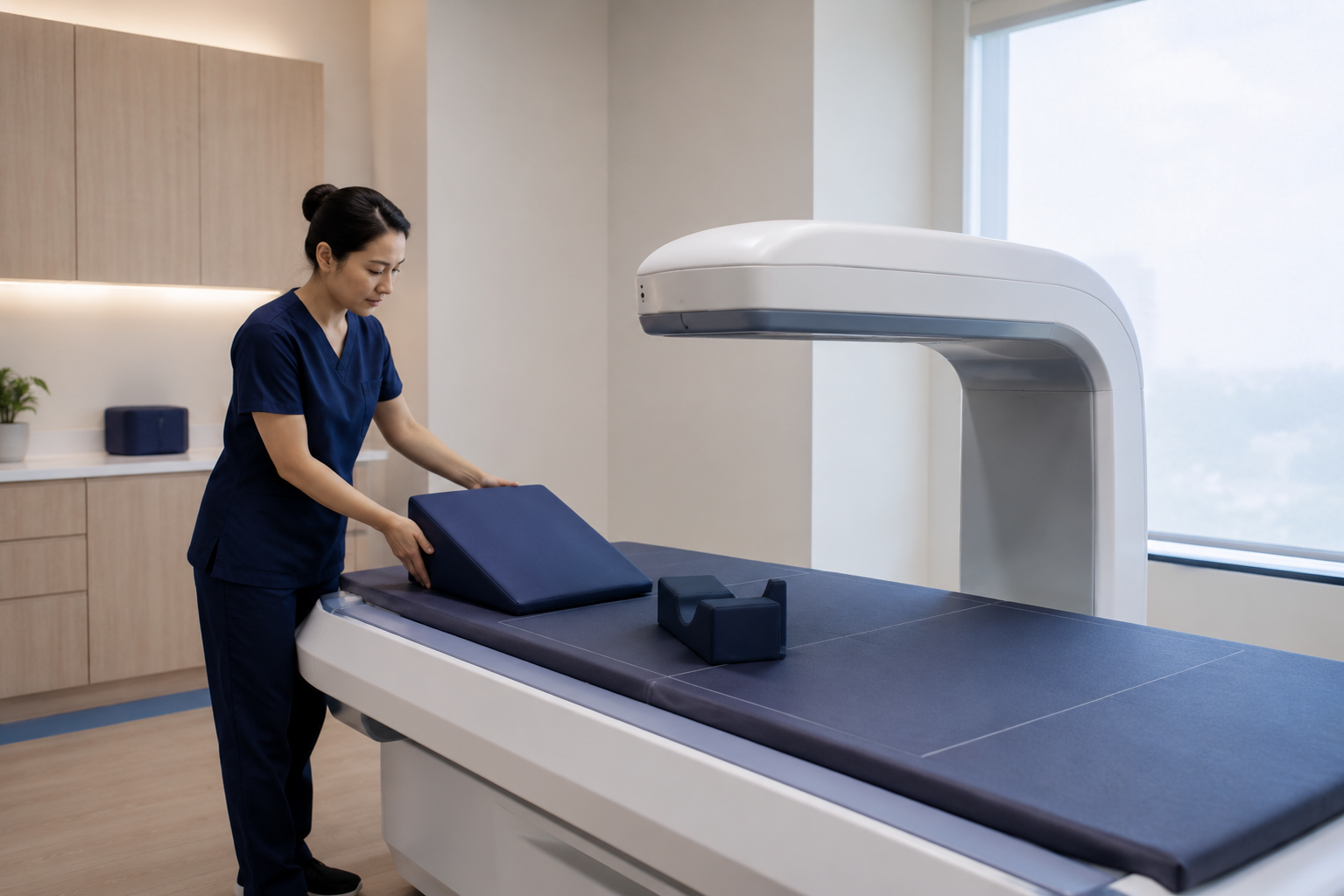
Bone Mineral Density (BMD) environment
06 Bone Mineral Density (BMD)

A short examination used to assess bone mineral density.
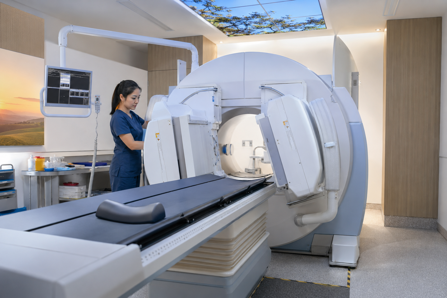
Nuclear Medicine environment
07 Nuclear Medicine

Confirmed services using approved radiopharmaceuticals should appear here.

Cyclotron & Radiopharmaceutical Production

Specialised imaging, closer to the point of care.

A cyclotron can produce selected short-lived radioisotopes for confirmed nuclear-medicine applications. Production, preparation and clinical use follow carefully controlled processes.
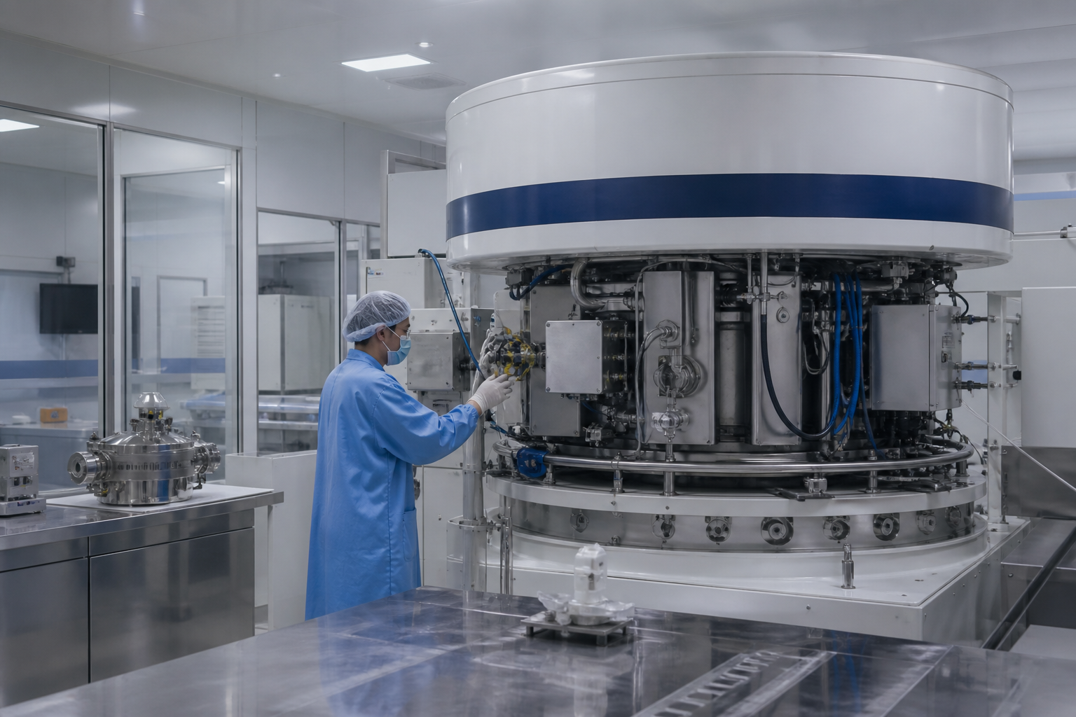
STATUS Planned capability
PURPOSE Support selected PET imaging
COMING SOON Cyclotron and radiopharmaceutical production facility

From production to clinical use

01 PRODUCE

Radioisotope production

Generate a selected medical radioisotope.

02 PREPARE

Radiopharmacy

Formulate and quality-check the radiopharmaceutical.

03 COORDINATE

Clinical scheduling

Coordinate time-sensitive preparation and use.

04 IMAGE

PET imaging

Use the tracer to answer a defined clinical question.

Technology in Clinical Use

Information and treatment, precisely connected.

Confirmed nuclear-medicine and radiotherapy services should be described according to their clinical and operational use.

Nuclear medicine imaging environment
TRACER-BASED SERVICES

Nuclear Medicine

Diagnostic or treatment services using approved radiopharmaceuticals, subject to confirmed clinical availability.
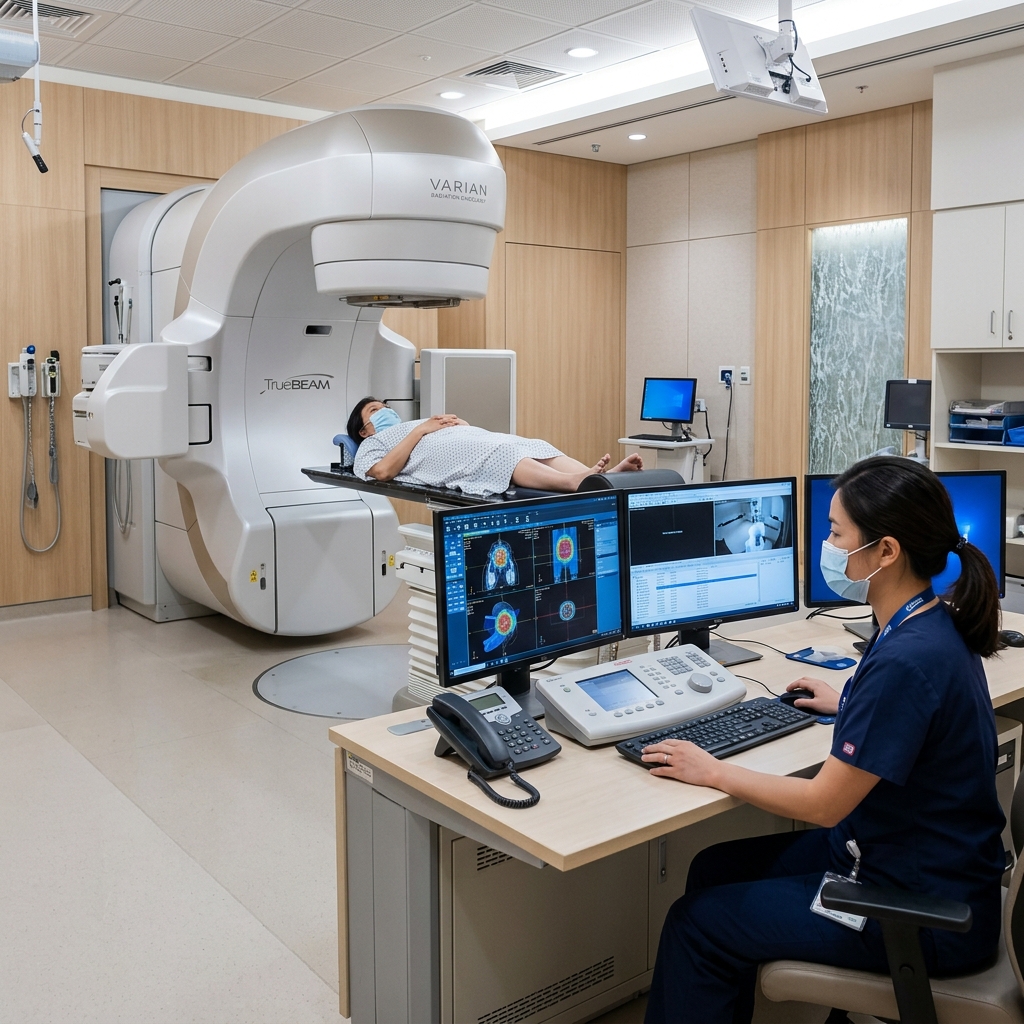
LINAC radiotherapy treatment room
PRECISION RADIOTHERAPY

Linear Accelerator (LINAC)

Technology designed to deliver carefully planned radiation therapy with precise positioning and treatment delivery.

On-Site Medical Laboratory & Pathology

Tissue, blood and molecular answers at clinical speed.

Imaging reveals where a tumour is located; our integrated clinical laboratory reveals its cellular and genetic composition. Same-day haematology, tumour marker profiling and rapid biopsy processing guide every step of precision cancer care.
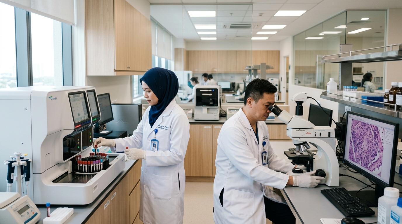
Medical laboratory scientists reviewing automated blood analysers and digital pathology
RAPID ON-SITE TURNAROUND

Immediate Laboratory Insights, Connected Directly to Treatment

Our on-site pathology and clinical laboratories are integrated with surgical theatres, daycare chemotherapy suites and imaging modalities—eliminating external courier delays and providing actionable answers at clinical speed.

ROUTINE BLOODS < 60 Mins Same-day pre-infusion clearance
INTRAOPERATIVE Real-Time Frozen section margin diagnosis
MDT TUMOUR BOARD Integrated Pathologist-led clinical review

Four Core Diagnostic Disciplines

Histology · Cytology · Haematology · Molecular Profiling
01
HISTOPATHOLOGY & CYTOLOGY

Tissue Architecture & Grading

Microscopic evaluation of needle biopsies and surgical resections, defining exact tumour lineage, histological grade, and margin clearance.

  • Biopsy & Surgical Resection Analysis
  • Intraoperative Frozen Section Assessment
  • Cytology & Fluid Cell-Block Evaluation
02
IMMUNOHISTOCHEMISTRY (IHC)

Biomarker & Receptor Profiling

Targeted antibody staining for hormone receptors and actionable surface proteins, determining suitability for targeted therapy and immunotherapy.

  • ER, PR & HER2 / neu Status for Breast Cancer
  • PD-L1 Expression for Immunotherapy
  • Diagnostic Subtype Differentiation Panels
03
HAEMATOLOGY & BIOCHEMISTRY

Pre-Treatment & Infusion Safety

Rapid automated screening of bone marrow reserve and metabolic organ function prior to chemotherapy, immunotherapy, or contrast-enhanced scans.

  • Full Blood Count & Absolute Neutrophil Count (ANC)
  • Renal Function & eGFR (Contrast Media Clearance)
  • Liver Function & Coagulation Profiles
04
MOLECULAR PATHOLOGY

Genomics & Tumour Markers

Detection of actionable somatic gene mutations and serial tracking of circulating serum tumour markers across treatment and follow-up.

  • EGFR, KRAS, BRAF & ALK Mutation Screening
  • Serial Tumour Markers (CEA, CA-125, PSA, AFP)
  • Microsatellite Instability (MSI / MMR) Testing
INTEGRATED CLINICAL WORKFLOW

How Laboratory Data Connects Directly to Patient Care

Accredited Clinical Quality
01 Specimen Collection

Gentle blood draw or tissue sample collection in our dedicated phlebotomy and clinical care suites.

02 Barcoded Chain-of-Custody

Automated electronic barcode tracking from bedside directly into the Laboratory Information System.

03 Automated Analysis & QA

Rigorous multi-level calibration, automated analysers, and pathologist validation ensure absolute accuracy.

04 Multidisciplinary Delivery

Real-time results transmitted to Oncologists and the Tumour Board (MDT) for immediate care decisions.

Preparing for Your Scan

Know what happens before you arrive.

Appointment and modality-specific instructions always take priority because preparation differs by examination and clinical need.

BEFORE YOUR VISIT

Be prepared for your appointment.

Keep your appointment letter, referral information, identification and relevant medical details together.

BRING WITH YOU Appointment letter Identification Medication information Relevant previous imaging
01
Referral reviewed The request and clinical question are checked so the examination can be planned.
02
Follow your instructions Preparation may differ for fasting, medicines, clothing, hydration or arrival time.
03
Confirm safety information Tell the team about relevant conditions, allergies, implants, devices and recent imaging.
04
Scan & specialist review The examination is completed and findings move through the referring care pathway.
Your appointment instructions take priority. Contact the imaging team if any preparation instruction is unclear or if your circumstances change.
Imaging Referral Information

The right information helps us prepare the right examination.

Patients and referring professionals can request examination-specific guidance and coordination information.

Speak with Our Team

Need help preparing for an examination?

Ask about an imaging appointment, referral requirements or examination-specific preparation.

Examination Guide

What patients may need to know